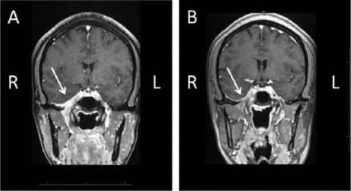

Anterolateral to the CPA, the internal auditory canal (IAC), a bony channel situated along the posterior face of the petrous bone, transmits the facial nerve from the CPA to the temporal bone and ultimately the face, and the vestibulocochlear nerve (VCN) from the cochlea and vestibular apparatus to the brainstem. The CPA contains cranial nerves V-VIII, the superior cerebellar artery (SCA), anterior inferior cerebellar artery (AICA), and draining veins. The cerebellopontine angle (CPA) is a cerebrospinal fluid-filled, triangular space located at the junction of the lateral pons and anterior cerebellum. She was not offered radiosurgery, and she elected conservative management. There was no evidence of a schwannoma on the repeat MRI. Repeat MRI demonstrated a loop of the anterior inferior cerebellar artery (AICA) compressing the vestibulocochlear nerve within the right IAC. She was originally diagnosed with a vestibular schwannoma on magnetic resonance imaging (MRI) and was referred to our institution for Gamma Knife radiosurgery. The current report represents an attempt to understand this clinical entity as discussed in the current literature.Ĭase summary: A 77-year-old female with a long history of progressive right-sided hearing loss and episodic vertigo developed unilateral right SSNHL, tinnitus, vertigo, and disequilibrium.

Underlying pathophysiologic factors surrounding microvascular compression of the vestibulocochlear nerve are poorly understood and make treatment recommendations, especially the option of microvascular decompression, difficult if not controversial.

Pneumo-CT is an effective means of diagnosing vascular loops and differentiating them from other lesions of the cerebellopontine angle.We present a patient with unilateral sudden sensorineural hearing loss (SSNHL) who was found to have a vascular loop in the ipsilateral internal auditory canal (IAC), and we review the literature regarding this association. Eighth nerve tumors and vascular loops produce similar symptoms, but a cochlear type of hearing loss with good speech discrimination and normal caloric testing should raise suspicion of a vascular loop. The wide range of audiometric and vestibular system test results probably reflects the complex interaction between the vascular loop and eighth cranial nerve, in which the loop exerts pressure on the nerve, and the nerve compromises inner ear circulation. Only one-third of the patients had abnormal caloric tests, but spontaneous nystagmus was detected in all but one of the patients by photoelectric nystagmography. Hearing losses ranged from mild to profound, and most were of a cochlear type with excellent speech discrimination. All patients were tumor suspects before CT because of unilateral (or asymmetric) tinnitus or hearing loss. In this study, we report the results of a uniform battery of audiometric and vestibular system test results administered to fifteen patients with prominent vascular loops in the internal auditory canal diagnosed by pneumo-CT. Previous reports have described pathologic anatomy, surgical approaches, and results of treatment. These vascular loops are suspected of causing hearing loss, tinnitus, and vertigo, and surgery has been advocated to separate the vascular loop from the eighth cranial nerve. Prominent loops of the anterior inferior cerebellar artery in the cerebellopontine angle are found frequently during anatomic studies of this region.


 0 kommentar(er)
